Mild Trigonocephaly - Autism - Ken
|
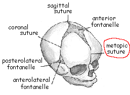
Please note that the text below represents my personal view and understanding. I am not a qualified medic but I have thoroughly studied the subject before deciding to have the surgical operation done for my 2 year old son. I hope you will find the information useful. What is mild trigonocephaly?
What is the relationship between mild trigonocelphaly and autism?Dr Shimoji told me that of the over 200 patients who had surgical operation since 1994, 80% of them has autism or has autistic tendencies. And over 90% of patients showed improvement after operation. If your child has mild trigonocelphaly and display any of the following symptoms - language delay, hyperactivity, motor dysfunction, impaired social interaction, head-banging, panic and irritability - then it is likely that mild trigonocelphaly is a contributing factor to the symptoms. Other symptoms related to the high intracranial pressure caused by mild trigonocelphaly include vomitting, morning headaches and squint. We do not know how many autistic or developmental delay children have mild trigonocelphaly or other forms of mild craniosynostosis but certianly it is not a small number. But having said that, I want to make it clear this is a very specific condition - not all autistic children have mild trigonocelphaly and therefore surgical operation will not help all cases of autism. How did mild trigonocelphaly contribute to autistic symptoms?Through two ways:
Note: (1) Functions of frontal lobe include intelligence, language, emotional control, fine motor movement, executive function, etc. (2) In Dr Shimoji's second paper all 56 patients have abnormally high intracranial pressure. Others have also reported high intracranial pressure in mild craniosynostosis. click here for a list of abstracts. (3) 80% of the over 200 patients has abnormal cerebral blood flow. Many published medical papers documented low or unbalanced cerebral blood flow in autistic individuals in particular the frontal lobe, click here for a list of abstracts. IMPORTANT NOTE: Trigonocephaly is one type of craniosynostosis. Other mild types of craniosynostosis may also produce the same effect. The two sites below will tell you more about the various types of craniosynostosis.What is the surgery and how does it help?The surgical operation is called decompressive cranioplasty. Normally the operation is carried out only for cosmetic purpose or emergency like car accident. The brain is completely untouched during the operation, only the bones are modified or rearranged. The operation achieve two things. First is that the widening of the forehead will allow the normal growth of frontal lobe. Click here to see the changes to Ken's forehead Click here to see the growth of the frontal lobe 6 months after operation New! Second is that the high pressure inside the head is released and this will lead to normal restoration of cerebral blood flow. As a result, many patients see improvements in the symptoms mentioned above. The surgery may not be a complete cure but it certainly helps a great deal in moderating the symptoms. Click here to find out how Ken is improving after operation. How do I know if my child has mild trigonocephaly?In most cases, mild trigonocephaly is palpable. Put your hand or fingers on the middle of the forehead, apply a bit of pressure and feel if there is a ridge (bone slightly sticking out) running anywhere between the top of nose to top of head. If there is a ridge, then your child has mild trigonocephaly. It may be easier to diagnosis when your child is asleep especially if your child is hyperactive or sensitive to touch. If you are not sure there is a ridge or not, or even if you are sure, you should go to see a Pediatric Neurosurgeon at a nearby hospital and asked for Three Dimensional Computed Tomography (3D-CT) scan of the child's head to be done to confirm the presence of the ridge. 3D-CT is the diagnosis test for mild trigonocephaly or other mild types of craniosynostosis. Standard CT or MRI can tell you if the frontal lobe is abnormally small but will NOT pick up mild trigonocephaly. Click here to see Ken's 3D-CT diagnostic test result. Apart from 3D-CT, are there any other diagnosis test?Yes, there are three other tests:
To find out moreClick below to download the following published papers by Dr Shimoji:
|
 Typical Trigonocephaly
 Ken's head before operation
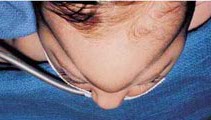
Very Mild trigonocelphaly
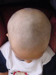
Ken's head before operation
Very Mild trigonocelphaly